- What should parents know about pediatric dental X rays?
- Are dental X rays safe for children?
- How much radiation do common pediatric dental X rays use?
- When do kids need dental X rays?
- Which types of dental images are used for children and what do they show?
- How do we make dental imaging comfortable for your child?
- What safety standards and regulations apply in California and the United States?
- How do dental X rays help with cavity detection and orthodontic assessment for children?
- What should you ask your pediatric dentist before your child gets X rays?
- Who are we and how do we provide pediatric dental imaging in Laguna Niguel and South Orange County?
- Frequently asked questions about pediatric dental X rays?
- References
What should parents know about pediatric dental X rays?
If you are a parent in Laguna Niguel or South Orange County, you probably want to know when dental X rays are necessary for your child and how safe they are. Pediatric dental X rays help us see areas that a visual exam cannot, such as between teeth, under fillings, along the roots, and around the developing jaws. They help us find early tooth decay, monitor growth, plan orthodontic care, check for extra or missing teeth, and evaluate injuries or infections. Professional organizations recommend that dentists use dental X rays only when they benefit the child and keep doses as low as reasonably achievable using modern digital systems and careful protocols [1] [2] [3].
We aim to be transparent about why an image is needed, what type is best, and how we keep exposure low. You can always ask us to explain the reason for any image and how it will guide your child’s care. The goal is to support healthy smiles while minimizing exposure with tailored decision making for each child [2] [3].
Are dental X rays safe for children?
When used judiciously, dental X rays for kids are considered a low exposure medical imaging exam. Modern digital sensors and thoughtful techniques reduce dose compared to older film. National and specialty guidelines emphasize using the lowest exposure settings that still provide a diagnostic image and taking images only when the expected benefits outweigh the small risks [1] [3] [4].
To put safety into context, the dose from common pediatric dental images is typically a fraction of what we receive from natural background radiation over an average day or two. For example, a set of bitewing images for cavity detection can be in the single digit micro sievert range with digital systems, while a digital panoramic image is commonly in the tens of micro sieverts. These ranges are reported across multiple studies and professional resources and are far below doses associated with deterministic effects. Even so, we always avoid unnecessary imaging and follow age appropriate protocols [4] [5] [6].
Protective measures can include thyroid shielding when it does not interfere with the exam, proper collimation and filtration, sensor holders that reduce retakes, and child specific exposure settings. Many professional bodies note that routine abdominal shielding is not generally necessary with modern equipment and techniques, yet patient centered shielding may be used when it adds comfort without compromising image quality. The key is a balanced, evidence informed approach for each child [1] [4].
How much radiation do common pediatric dental X rays use?
Parents often ask for numbers. While the exact dose depends on the child’s size, the machine, and technique, researchers and professional bodies provide useful ranges for comparison. Below are practical perspectives drawn from peer reviewed literature and expert guidelines [4] [5] [6].
What are typical effective dose ranges for dental images in children?
- Digital bitewing images for cavity detection These commonly measure in the low single digit micro sieverts per image, with small total values for a standard set, especially when rectangular collimation and high sensitivity sensors are used [5] [6].
- Periapical images to evaluate specific teeth These are generally similar to bitewings on a per image basis when using digital sensors and proper collimation [5] [6].
- Digital panoramic image Typical pediatric values are commonly in the tens of micro sieverts, varying with equipment and protocols. Panoramic imaging surveys both jaws and is not usually taken at every visit [5].
- Cephalometric image for orthodontic assessment This lateral skull image often measures in the low single digit micro sieverts when using contemporary digital systems [5].
- Dental CBCT limited field of view for a focused question Pediatric doses vary widely depending on field of view and exposure settings, from values comparable to or modestly above panoramic imaging up to higher ranges when larger fields or higher settings are used. CBCT is reserved for cases where added 3D information is necessary and conventional images are insufficient [4] [6].
How does dental radiation compare to everyday background exposure?
Everyone receives background radiation from natural sources every day. The combined dose from a typical set of digital bitewings is often in the range of a day or a few days of natural background exposure, while a digital panoramic image may be in the range of several days. These comparisons help put small medical exposures into perspective and are used by professional organizations in risk communication with families [4] [5].
Why do digital systems reduce dose compared with film?
Digital sensors are more sensitive than traditional film. They allow lower exposure settings while maintaining diagnostic quality, especially when paired with rectangular collimation and filtration. Digital workflows also reduce retakes by providing instant feedback to the dental team and facilitating quality control, which helps keep total exposure low for children [1] [5].
When do kids need dental X rays?
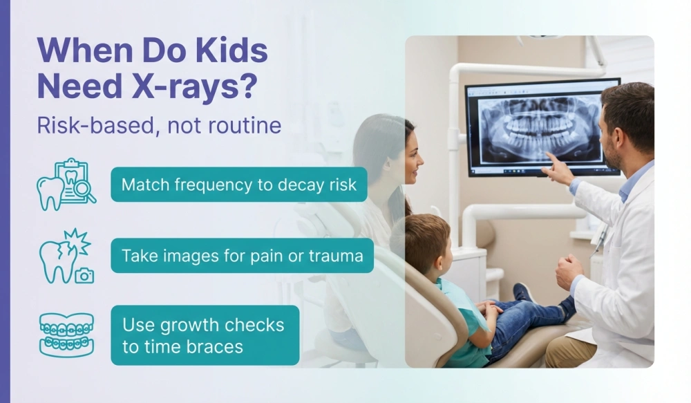
Children need dental X rays based on individualized risk and specific clinical questions. There is no one size fits all schedule. Dentists consider the child’s age, oral hygiene, diet, fluoride exposure, history of cavities, tooth development, and symptoms. Professional guidelines from the American Dental Association and American Academy of Pediatric Dentistry focus on risk based intervals and selection criteria rather than routine imaging at every visit [2] [3].
How does risk level guide the frequency of bitewing images?
- Low risk children with no clinical decay Bitewing images may be taken at longer intervals to screen for hidden decay between teeth. The time interval is typically extended when risk is low and teeth are spaced, especially in younger children [2] [3].
- Higher risk children with past or active decay Bitewing images are recommended more frequently to monitor for new or progressing decay between teeth, which is an area not visible in a simple visual exam [2] [3].
- Children with orthodontic appliances Risk may increase around brackets and bands, so your dentist may recommend images at shorter intervals while appliances are in place, paired with added prevention strategies [3].
What signs or symptoms indicate images are needed sooner?
- Localized pain or swelling Targeted periapical images help evaluate possible infection, injury, or deep decay that may not be visible otherwise [3].
- Trauma to the teeth or jaw Images help assess root fractures, displacement, or effects on developing permanent teeth after a fall or sports injury [3].
- Delayed eruption or missing teeth Panoramic or targeted images can identify impacted, extra, or missing teeth and guide timing of orthodontic evaluations [3].
- Monitoring growth and development At certain stages, orthodontic assessment images help the dentist evaluate jaw relationships and growth patterns for planning interceptive care [3].
Which types of dental images are used for children and what do they show?
Different images answer different questions. Using the right view at the right time keeps exposure low and yields the answers we need to guide care [2] [3].
How do digital bitewing images help detect cavities between teeth?
Digital bitewing images show the crowns of the upper and lower teeth in one area and are highly useful for cavity detection for children where teeth touch. They help us find early decay and catch issues before they cause pain, which can reduce the need for more extensive treatment later. Because children often develop decay between molars, these images are a mainstay of preventive care when clinically indicated [3] [5].
What questions do periapical images answer around the root and supporting bone?
Periapical images show the entire tooth from crown to root tip and the surrounding bone. They are useful for evaluating deep cavities, dental abscesses, root development, trauma, or issues around a specific tooth. For a child with a persistent toothache, this focused image can be the most efficient way to identify the cause [3].
When is a panoramic image helpful for children?
A panoramic image surveys the entire upper and lower jaws, sinuses, and developing teeth in one pass. It can be helpful for growth and development checks, timing orthodontic referrals, evaluating extra or missing teeth, and screening for cysts or jaw problems. It is not needed at every visit and is ordered when it will change care decisions or timing [3] [5].
Why might a cephalometric image be used in orthodontic planning?
A cephalometric image is a side view of the face and skull used to measure jaw relationships, growth direction, and airway dimensions. Orthodontists use these measurements to plan braces or early growth guidance. With digital systems, this image typically carries a low dose while providing valuable planning information [5].
When is dental CBCT appropriate for a child?
CBCT provides three dimensional information and is reserved for specific questions that cannot be answered with two dimensional images, such as exact position of impacted teeth, evaluation of pathology, or detailed assessment before certain procedures. When CBCT is indicated, we choose the smallest field of view and lowest exposure that answers the question, in line with expert guidance for pediatric imaging [4] [6].
How do we make dental imaging comfortable for your child?
Comfort matters as much as clarity. A relaxed child moves less and that reduces the chance of retakes. Here is how we make imaging easier.
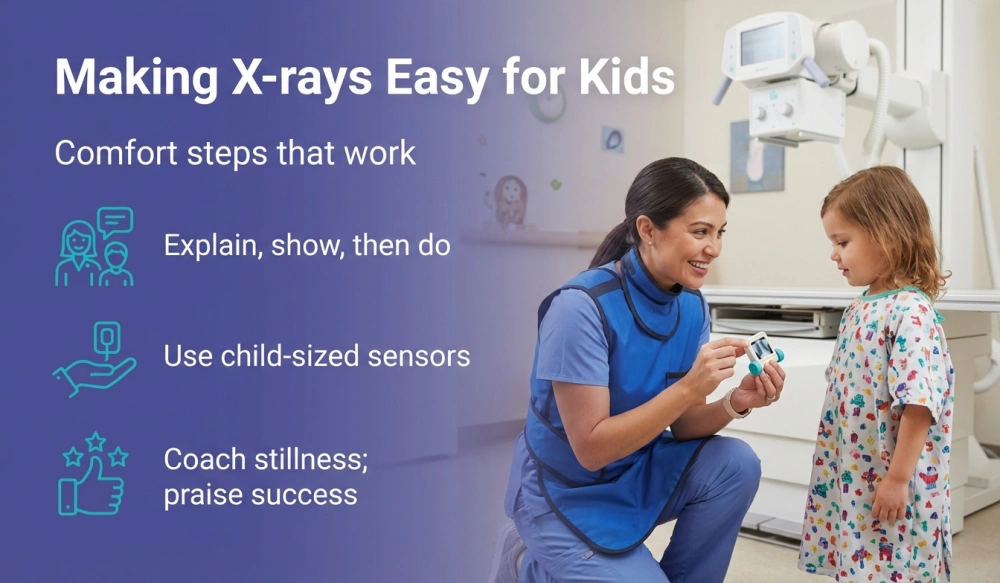
- Tell show do We explain what will happen in simple words, show the sensor and camera, and then gently take the image so children know what to expect.
- Child sized sensors and holders We use small, flexible tools and positioners so sensors feel more comfortable in little mouths.
- Practice breaths and stillness We coach kids to be still for a short count and celebrate their success right after the click.
- Topical comfort aids When appropriate, a small amount of topical gel can make the sensor edges more comfortable.
- Parent presence Parents are welcome to be nearby when it helps the child feel calm, following safety rules for the imaging room.
What safety standards and regulations apply in California and the United States?
Dental offices in California follow both federal guidance and state specific rules for X ray equipment, operator training, and patient protection.
- Federal guidance The FDA and professional groups publish recommendations for prescribing dental radiographs based on patient selection criteria and for using current technology to limit exposure. These recommendations support clinical decision making and quality assurance in dental radiography [1] [2].
- State registration and compliance In California, dental X ray machines must be registered with the California Department of Public Health Radiologic Health Branch. Practices must follow state rules for installation, inspection, and safe operation of radiation producing equipment [7] [8].
- Office protocols and ALARA We implement office policies that reflect ALARA principles, now often expressed as ALADA for as low as diagnostically acceptable. This means tailoring exposure to the child’s size and the clinical need and avoiding retakes through careful positioning and training [3] [4].
- Shielding and collimation practices We use collimation to focus the beam and child appropriate exposures. Patient shielding such as a thyroid collar may be used when it does not interfere with the exam and aligns with contemporary guidance from expert bodies. The emphasis is on technique first and judicious shielding second for pediatric comfort and safety [1] [4].
If you have questions about how we implement these guidelines in Laguna Niguel and across South Orange County, we are happy to walk you through our checklist and demonstrate our equipment settings at your child’s visit.
How do dental X rays help with cavity detection and orthodontic assessment for children?
Dental X rays allow us to see problems early, often before a child feels symptoms. Early detection usually means simpler care.
- Cavity detection for children Bitewings reveal decay between teeth where toothbrush bristles cannot reach. That is one reason many school age children benefit from periodic bitewings at intervals matched to their risk. Catching early decay pairs well with fluoride, sealants, and home care to avoid bigger issues later [3] [9].
- Monitoring growth and development Panoramic and cephalometric images help time orthodontic evaluations, check for crowding, and evaluate jaw relationships. These insights support interceptive steps like space guidance or timing extractions when recommended by the orthodontic team [3].
- Orthodontic assessment X rays When a child is entering mixed dentition, panoramic and cephalometric images provide a broad view. If an impacted canine position is uncertain or root resorption is suspected, a limited field CBCT may be considered when conventional images do not answer the question [4] [6].
- Injury and infection evaluations Focused periapical images help diagnose abscesses or trauma effects so we can treat promptly and protect developing permanent teeth [3].
What should you ask your pediatric dentist before your child gets X rays?
- Why is this image needed today Ask what clinical question the image will answer and how it could change care decisions [2] [3].
- Which type of image is best Discuss whether bitewing, periapical, panoramic, cephalometric, or CBCT is most appropriate for the question at hand [3] [4].
- How is exposure kept low Ask about digital sensors, rectangular collimation, child specific settings, and steps to minimize retakes [1] [5].
- Will shielding be used If you want a thyroid collar for your child, ask whether it can be used without interfering with the image and how it fits within current guidelines [1] [4].
- How often will images be repeated Request a risk based plan tailored to your child rather than a fixed schedule, and ask how home care and diet can reduce the need for frequent imaging [2] [3].
- How are images stored and shared Confirm that digital images are securely archived and can be shared with your orthodontist or other providers as needed.
Who are we and how do we provide pediatric dental imaging in Laguna Niguel and South Orange County?
Kidz Modern Dentistry provides pediatric dental care for families across Laguna Niguel, Laguna Hills, Aliso Viejo, Laguna Beach, Mission Viejo, and San Juan Capistrano. Our team focuses on education first. When an image is recommended, we explain the clinical reason, the type of view, and how it will impact your child’s treatment plan. We use child specific exposure settings and modern digital sensors to obtain clear images with low dose techniques aligned with current guidance [1] [3].
We invest in streamlined digital imaging systems, quality assurance protocols, and staff training that emphasize accurate positioning and minimal retakes. Our approach includes standardized exposure charts by size and dentition stage, regular equipment calibration checks, and peer review of image quality so that one clear image answers the clinical question whenever possible [5].
Our practice offers same day help for urgent concerns, kid friendly sedation when appropriate, and comprehensive pediatric care under one roof. We coordinate closely with local orthodontists for growth monitoring and interceptive planning. For example, a panoramic image may help time an orthodontic referral, while bitewings monitor cavity risk during orthodontic treatment. When a problem calls for three dimensional detail such as the exact position of an impacted canine, we coordinate a limited field CBCT at the lowest settings that answer the question, in line with pediatric guidelines [4] [6].
Our service area includes Laguna Niguel and the communities of South Orange County. Parents looking for kids dental X rays near me often want to know how we keep care gentle. We emphasize clear communication, comfort coaching, and step by step demonstrations for young patients. In addition, we use pediatric sized holders and sensors to make the process easier for small mouths.
We also support collaborative care by securely sharing images with your child’s medical or dental specialists. This avoids duplicate imaging and keeps your child’s overall exposure low. When we say we focus on comfort and clarity, we mean that every step from scheduling to follow up is designed to make visits smooth for your family. If you are comparing options for children’s dental imaging in Laguna Niguel and South Orange County, we welcome your questions about our protocols and equipment.
For families who prefer a practice experienced in modern imaging, we provide Pediatric digital X-rays in Laguna Niguel using digital sensors, rectangular collimation, and child centered workflows that align with current recommendations. To talk with a pediatric dentist about your child’s needs, call us at +1 818 260 0016 and we will be happy to help you plan next steps.
Frequently asked questions about pediatric dental X rays?
Do baby teeth ever need dental X rays?
Yes, primary teeth can benefit from images when the dentist suspects hidden decay, checks development, or evaluates injury. Images are prescribed only when the expected benefit outweighs the small exposure, and techniques are adjusted for young children to keep dose low [2] [3].
How often should children without cavities have bitewing images?
There is no fixed interval for every child. For those at low risk with no signs of decay, guidelines support longer intervals between bitewing sets. The dentist will consider your child’s spacing, diet, fluoride exposure, and oral hygiene to set an interval that fits their risk level [2] [3].
Are dental X rays safe for children with special health care needs?
Yes when they are clinically indicated and tailored to the child’s needs. Techniques such as shorter exposure times, positioning aids, caregiver support, and thoughtful shielding can help complete the study comfortably while keeping dose low. The dentist weighs risks and benefits for each child and coordinates with the medical team as needed [3] [4].
What if my child cannot tolerate an intraoral sensor comfortably?
We begin with smaller sensors, practice placement, and gentle coaching. If an intraoral view is not possible, the dentist may consider alternative views such as a panoramic image for a broad overview or may defer imaging until it becomes necessary and feasible. The decision depends on the clinical question and the child’s comfort [3].
Is a thyroid collar used during dental X rays for kids?
Many practices use a thyroid collar for children when it does not interfere with the image. Contemporary guidance emphasizes optimized technique and collimation as primary protection. A collar is reasonable when it fits the exam and does not cover the area of interest. Your dentist can explain how shielding is chosen for each view [1] [4].
Does my child need a panoramic image every year?
No. A panoramic image is recommended when it answers a specific growth, development, or diagnostic question that cannot be answered another way. The timing is individualized and not tied to a yearly schedule [3].
When is CBCT considered for a child and how is dose managed?
CBCT is considered when three dimensional detail is necessary to plan care and two dimensional images are insufficient. When used, pediatric protocols select the smallest field of view and the lowest exposure that still provides diagnostic quality, following expert guidance for children [4] [6].
Can dental X rays reveal orthodontic problems early?
Yes. Panoramic and cephalometric images can show crowding, missing or extra teeth, and jaw relationships. These findings help time orthodontic evaluations and guide referrals for interceptive orthodontics when appropriate [3].
Will dental X rays be covered by insurance?
Many plans include dental imaging as part of preventive and diagnostic services, but coverage varies widely. Your plan documents or insurer can confirm what is included and how often images are covered. Our team can provide fee information and help you submit claims.
How are my child’s images protected and shared when needed?
Digital images are stored in secure systems and shared with other providers only with your consent for care coordination. Secure sharing helps avoid unnecessary repeat imaging and keeps total exposure low.
What can I do at home to reduce the need for frequent images?
Strong prevention lowers cavity risk and often reduces the need for frequent bitewings. Focus on daily brushing with fluoride toothpaste, flossing where teeth touch, a balanced diet low in sugary snacks and drinks, and regular checkups. Community water fluoridation and professional fluoride or sealant treatments are additional protective steps that reduce decay risk [9].
Are dental X rays needed after a sports injury to the mouth?
Often yes. Targeted images help check for root fractures, displacement, or injury to the developing permanent teeth. Timely imaging and evaluation support appropriate treatment and follow up [3].
How do low dose digital X rays fit into care for kids with braces?
Orthodontic appliances can trap plaque and raise cavity risk. Risk based bitewings may be recommended during treatment, and panoramic or cephalometric images support planning and progress checks. With digital sensors and child specific settings, the dose remains low and images are timed to clinical need [3] [5].
References
- Dental Radiography Guidance for Patients and Providers – U.S. Food and Drug Administration
- Dental Radiographic Examinations Recommendations for Patient Selection and Limiting Radiation Exposure – American Dental Association
- Prescribing Dental Radiographs for Infants, Children, Adolescents, and Individuals With Special Health Care Needs – American Academy of Pediatric Dentistry
- Dental Imaging Resources for Children – Image Gently Alliance
- Understanding Dose from Dental X Rays – Health Physics Society
- Radiation Protection No. 172 Cone Beam CT for Dental and Maxillofacial Radiology Guidelines – European Commission SEDENTEXCT
- Registration of Radiation Producing Machines – California Department of Public Health Radiologic Health Branch
- Radiologic Health Branch Laws and Regulations – California Department of Public Health
- Children’s Oral Health Tooth Decay Facts – Centers for Disease Control and Prevention
- X Rays and Dental Care Information – National Institute of Dental and Craniofacial Research